Je confirme être un professionnel de santé*

Valider

* Conformément à la loi française de décembre 2011 relative au renforcement de la sécurité sanitaire et à ses décrets d'application de mai 2012, « les sections des sites internet réservées aux seuls professionnels de santé doivent être, à minima, accessibles après une page d'engagement de l'internaute, certifiant qu'il est un professionnel de santé. »

Informations sur les cookies

Les cookies sont des petits fichiers installés sur votre ordinateur ou votre smartphone. Ils nous permettent de stocker des informations relatives à votre navigation sur notre site.

En savoir plus Accepter les cookies

Politique de confidentialité

Accepter les cookies Fermer

Informations sur les cookies

Olea Medical utilise trois types de cookies :

  • Les cookies de session ou de préférences qui sont indispensables à la navigation et au bon fonctionnement du site
  • Les cookies de mesure d’audience et autres traceurs servant à établir les statistiques d’audience du site

Ces cookies sont paramétrés selon les critères d’exemption du consentement tels que définis par la CNIL

Nom du cookieTypeDescription
Nécessaire à la fourniture du serviceMémorise l'attestation obligatoire "professionnel de santé"
Nécessaire à la fourniture du serviceStocke le consentement pour chaque cookie de type “traceur”.
Nécessaire à la fourniture du serviceStocke l'authentification nécessaire au téléchargement des cases reports
Nécessaire à la fourniture du serviceNécessaire au fonctionnement du site. Permet de savoir si les cookies sont activés.
Nécessaire à la fourniture du serviceStocke la langue courante
Authentification clientEspace client uniquement. Ce cookie ne sera pas installé si vous ne vous connectez pas à votre compte.
Nécessaire à la fourniture du serviceEspace client uniquement.
Nécessaire à la fourniture du serviceCookie CloudFlare nécessaire à la protection des bots
Mesure d'audienceCookie principal permettant le suivi des visiteurs via le service hubspot
Mesure d'audienceCookie permettant le suivi des sessions via le service hubspot
Mesure d'audienceUtile au suivi suivi des sessions via le service hubspot
Mesure d'audienceCookie lié au service hubspot
Mesure d'audienceSuivi des visites via google analytics

Amide Proton Transfer weighted imaging reveals a rolandic tumor in a pediatric patient

Nathalie BODDAERT, MD, PhD, Pediatric Neuroradiologist, Necker-Enfants Malades Hospital, AP-HP, Pediatric Radiology Department, Université Paris, Paris, France
Volodia DANGOULOFF-ROS,MD, PhD, Pediatric Neuroradiologist, Imagine Institute, INSERM U1163, Université Paris cité, F-75015, Paris, France

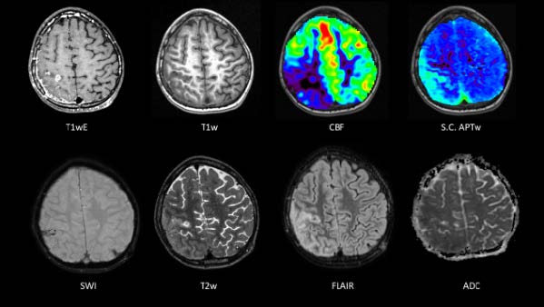
Introduction Discovery of a right parietal lesion in a five-year-old child with local meningeal extension following a convulsive seizure. Medical history The young patient presented a first hyperthermic convulsive seizure. One month later, he presented another tonic-clonic episode with left hemicorporeal seizure lasting less than 5 minutes. Another month later, he had a tonic-clonic seizure again, without fever but complicated by status epilepticus. Afterwards, a CT scan showed a right parietal lesion, and he was transferred to the Necker- Enfants Malades hospital for a brain and spinal-cord MRI. The spinal-cord MRI did not reveal any metastases. A lesionectomy was performed. A postoperative remnant and a local meningeal infiltration were shown on brain MRI. Anatomical pathology indicated a right parietal atypical teratoid rhabdoid tumor (ATRT), which is an embryonal tumor proliferation, INI1 deficient. The young patient had a first course of chemotherapy with doxorubicin followed by radiotherapy (from 3 months to 4 months after surgery). Image acquisition and analysis Examinations were acquired on 3T MRI (GE Milwaukee, MR750W) at different timelines: pre-operative, post-operative (48 hours, 6 weeks and 10 weeks) after neurosurgery. For each timeline, 3D gradient echo (GRE) T1-weighted, 3D fast spin echo (FSE) T2-weighted, 3D FSE FLAIR, Diffusion-weighted (12 directions), Susceptibility Weighted Imaging (SWI) and post-contrast 3D FSE T1-weighted sequences were acquired. Pseudo continuous arterial spin labelling (pCASL) sequences were performed to measure Cerebral Blood Flow (CBF) in post-operative MRI. 3D FSE Amide Proton Transfer weighted (APTw) sequence was performed in post-operative 6-week timeline in the framework of a clinical research study (see Additional Information). Apparent Diffusion Coefficient (ADC) and CBF maps were computed by the scanner software, while Spillover-corrected APTw (S.C. APTw) maps were processed in Olea Sphere 3.0, to correct for movement, noise, field inhomogeneities and artefacts from fluid compartments. Examinations were reviewed in consensus by two senior pediatric neuroradiologists. Follow-up 6 weeks after the surgery. The MRI protocol showed persistence of leptomeningeal contrast enhancement. A contrast-enhanced lesion with no diffusion restriction and with a decreased CBF was observed in the right rolandic sulcus. FLAIR hypersignal was persistent in the operated area and at the rolandic level. However, at this rolandic level, the APTw map revealed a marked relative increase in signal intensity. Mobile proteins and peptides are the main contributors of APTw contrast, known to increase in neoplastic tissue. Therefore, S.C. APTw suspected a rolandic tumor, whereas ADC and CBF maps missed revealing such finding (Figure 1).
Cr 05 23 Cest Pediatric Fr Olea Image 1
   Figure 1 MR sequences at 6 weeks. S.C. APTw = Spillover-corrected APTw. CBF = ASL-CBF. T1wE = T1w after Gadolinium-based agent injection

10 weeks after the surgery. The young patient was clinically stable. Multimodal MRI repeatedly showed the persistence of leptomeningeal contrast (Figure 2).
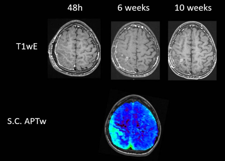
No increase in CBF nor decreased ADC signal intensity was shown in the rolandic region. Conversely, the contrast enhancement on T1wE was increased in favor of tumor progression not totally removed by the surgeon, at the exact location where APTw map revealed a large increase in amide signal intensity 4 weeks before (Figure 3). 

Cr 05 23 Cest Pediatric Fr Olea 2
                                    Figure 2 MR sequences at 10 weeks. CBF = ASL-CBF. T1wE = T1w after Gadolinium-based agent injection
Cr 05 23 Cest Pediatric Fr Olea 3
                           Figure 3 : T1wE enhancement follow-up, in favor of tumor progression at 10 weeks, as shown by S.C. APTw at 6 weeks

Conclusion
Amide proton transfer weighted MRI was found to be a non-invasive and useful biomarker in predicting the outcome of this brain condition. More frequent use in pediatric clinical routine could help us understand its full predictive potential.

Additional information and Acknowledgements
Local Institutional Review Board authorization was granted (EDRACT 2014-A00541–46). We thank Dr. Thomas Blauwblomme, Dr. Christelle Dufour, Dr. Pascale VARLET, Julie Poujol PhD, Stefano Casagranda PhD, for the material and support for this Case Report.

Pour en savoir plus sur notre solution CEST