Cookie information

Cookies are small files installed on your computer or smartphone. They allow us to store information about your navigation on our site.

Choose cookies Accept all cookies

Privacy Policy

Accept all cookies close

Cookie information

Olea Medical uses three types of cookies:

  • Session or preference cookies, which are essential for navigation and the good functioning of the site
  • Audience measurement cookies and other tracers used to establish site audience statistics

These cookies are configured according to the criteria for exemption from consent as defined by the CNIL

CookieTypeDescription
Service supplyStores consent for each "tracker" type cookie.
Service supplyStores authentication related to case report downloads
User authenticationCustomer area only. This cookie will not be set if you do not log in to your account.
Service supplyStores the current language
TrackingGoogle analytics

Neuroimaging Neuro-oncology MRI solution
  • MRI

The complete MRI solution for improved analysis and optimum patient management

Optimized workflow and treatment monitoring

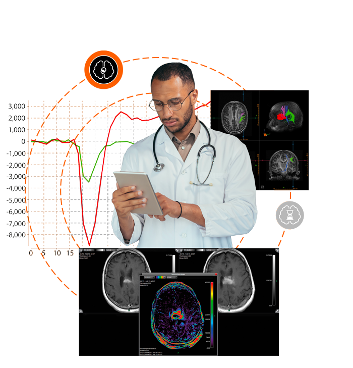
By offering a full range of innovative functions, our neuro-oncology solution allows you to procure more precise results and to interpret the images with greater clarity in order to provide patients with more effective treatments.

This multi-vendor solution offers a complete range of innovative functions, guaranteeing results of unequalled precision and greater clarity when interpreting the images, thus helping with patient care.

Fluidly integrated into your workflow, the solution has been designed to assist you at every stage in your decision-making process. It is compatible with most constructors and provides essential information for numerous neurological diseases, as well as a more precise vision of the treatment response and the development of the disease.

Our neuro-oncology solution is combined with neuronavigation systems, thus making the planning and navigation easier during neurosurgery. In addition, it improves the pre- and post-operative follow-up by merging the traditional maps with the tractography results. You therefore benefit from a precise view of the cerebral structures and their connections and can optimize your surgeries.

Solution imagerie en neurologie

An innovative suite for unparalleled diagnostics and treatment follow-up

With expertise supported by over one hundred clinical publications on brain tumors, we make the most advanced tools for cutting-edge MRI analyses available to radiologists, surgeons, and oncologists.

Our suite of neuro-oncology solutions offers you essential quantitative information at each crucial step in the patient’s pathway, while simplifying and speeding up the comparison with previous examinations.

  • A simplified decision-making process
  • Precise assessment
  • Guaranteed qualified thanks to constant results
Neurology booklet Download
  • Olea case report: Brain Tumor: Low Grade & High Grade Gliomas
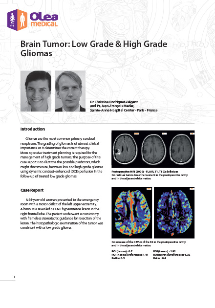
    Brain Tumor: Low Grade & High Grade Gliomas
    Gliomas are the most common primary cerebral neoplasms. The grading of gliomas is of utmost clinical importance as it determines the correct therapy. More aggressive treatment planning is required for the management of high-grade tumors. The purpose of this case report is to illustrate the possible predictors which might discriminate between low-and-high grade gliomas using dynamic contrast-enhanced (DCE) perfusion in the follow-up of treated low grade gliomas.
  • Olea case report: Brain Tumor: Glioblastoma with Oligodendroglial Component
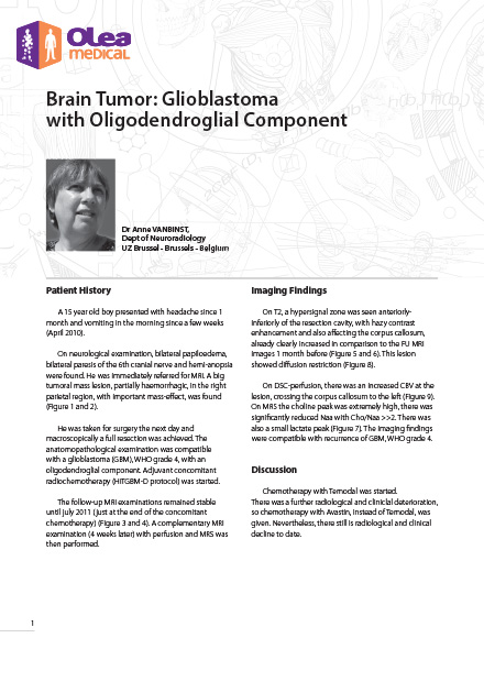
    Brain Tumor: Glioblastoma with Oligodendroglial Component
    A 15-year-old boy presented with headache for 1 month and vomiting in the morning for a few weeks. On neurological examination, bilateral papiloedema, bilateral paresis of the 6th cranial nerve and hemianopia were found.
  • Olea case report: Brain Tumor: Low-Grade Glioma
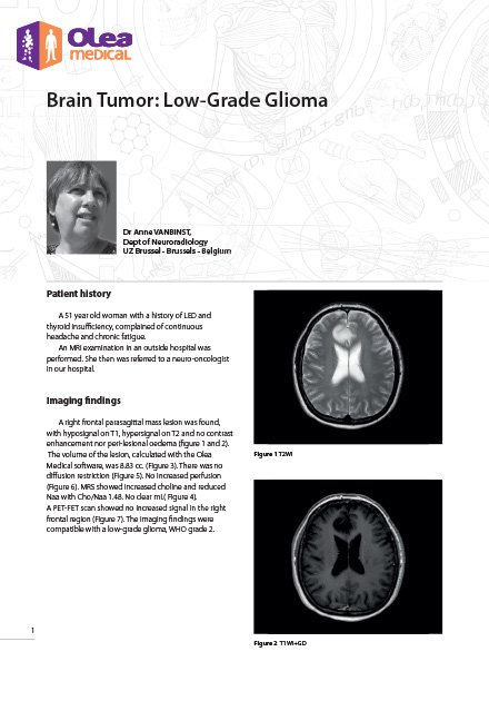
    Brain Tumor: Low-Grade Glioma
    A 51-year-old woman with a history of LED and thyroid insufficiency, complained of continuous headache and chronic fatigue. An MRI examination in an outside hospital was performed. She then was referred to a neuro-oncologist.

3 figures to remember

On average

person will develop a neurological disorder in their life (1)
1/ 1

Up to

patients with cancer will develop cerebral metastases (in the USA) (2)
0 %

On average

is the life expectancy lost due to brain tumors, the highest of all cancers
0 years

Overview of the main functions

Calculating the ADC map
Calculating a complete set of T2* perfusion maps (DSC): CBV (Cerebral Blood Volume), K2 (qualitative permeability map for assessing the integrity of the BBB), corrected CBV, CBF, MTT, TTP, Tmax and tMIP
Automated or manual selection of the arterial input functions (AIF)
Multi-parameter ROI/VOI/3D analysis
Generation of T1 perfusion maps (DCE): semi-quantitative Kep, Ktrans, Ve and Vp maps and optimized qualitative TME (Time to Maximum Enhancement), AUC (Area Under Curve), Wash-in, Wash-out, Peak, Peak Enhancement, SER (Signal Enhancement Ratio) and Curve washout maps
MRI perfusion technique without Arterial Spin Labeling (ASL) contrast: mapping weighted for perfusion and blood flow
fMRI: maps with activations zones in the three planes - Laterality index
Compatibility with neuronavigation systems
CT perfusion: 4 deconvolution methods: sSVD, cSVD, oSVD and patented Bayesian
Option to establish longitudinal ratios: temporal reset of sequences - Temporal subtraction - Development as a percentage of the volume
Generation of reports with summarized results
Option of merging maps
Integration with multiple PACS in order to share results
Multi b: Diffusion, IVIM, Kurtosis, Full Kurtosis (DWI/IVIM/Kurtosis - research)  Optional
APT CEST  Optional

Complete your neuro-oncology offer

Discover our emergency solutions for managing strokes.

The clinical and research institutes at our side

They put their trust in us

Logo CHU Bordeaux
UCLA
Hunimed
Logo I.R.C.C.S. Ospedale San Raffaele
Logo Mayo Clinic
Logo Fondazione Mondino

Give it a go!

Request a demo or contact our sales team

Renseignez votre email dans le formulaire ci-dessous afin de pouvoir télécharger les case reports sur le site olea-medical