Cookie information

Cookies are small files installed on your computer or smartphone. They allow us to store information about your navigation on our site.

Choose cookies Accept all cookies

Privacy Policy

Accept all cookies close

Cookie information

Olea Medical uses three types of cookies:

  • Session or preference cookies, which are essential for navigation and the good functioning of the site
  • Audience measurement cookies and other tracers used to establish site audience statistics

These cookies are configured according to the criteria for exemption from consent as defined by the CNIL

CookieTypeDescription
Service supplyStores consent for each "tracker" type cookie.
Service supplyStores authentication related to case report downloads
User authenticationCustomer area only. This cookie will not be set if you do not log in to your account.
Service supplyStores the current language
TrackingGoogle analytics

Oncology imaging
  • MRI

Your complete solution for oncology imaging

Full assessment within the oncology treatment continuum

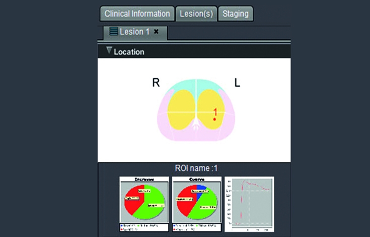
Our solution gives access to the latest tools and applications for image analysis of a wide range of organs: breast, female pelvis, prostate, and rectum. It is helpful both for routine clinical use and for further study, by quickly calculating all the maps and measurements expected in the target regions of interest. Our solution also provides access to standardized reports based on the latest versions of the international BI-RADS® and PI-RADS® classifications for breast and prostate studies.

Optimized solutions to tackle the complexities of oncology imaging

Our technology allows for quicker characterization of lesions, increased confidence in the treatment strategy and better assessment of the response to treatment.

  • Intuitive multi-parameter user interface
  • Standardized reports
  • ADC and diffusion mapping
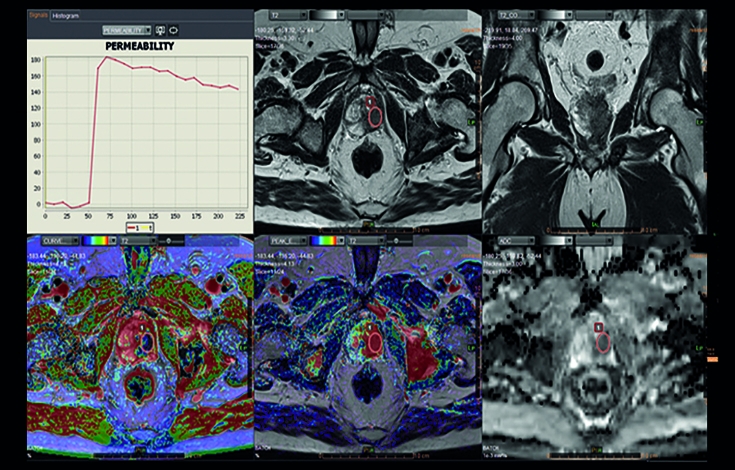
  • Graphic evaluation of the kinetics
  • Calculation of Electronic b maps(proprietary)
Neurology Solutions Download
  • Olea case report: Metastatic Pancreatic Cancer
    Metastatic Pancreatic Cancer
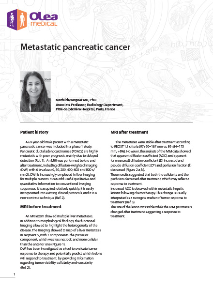
    A 69-year-old male patient with a metastatic pancreatic cancer was included in a phase 1 study. Pancreatic ductal adenocarcinomas (PDACs) are highly metastatic with poor prognosis, mainly due to delayed detection (Ref. 1). An MRI was performed before and after treatment, including diffusion-weighted imaging (DWI) with 6 b-values (0, 50, 200, 400, 600 and 800 s/ mm2).
  • Olea case report: Lower Limb Sarcoma
    Lower Limb Sarcoma
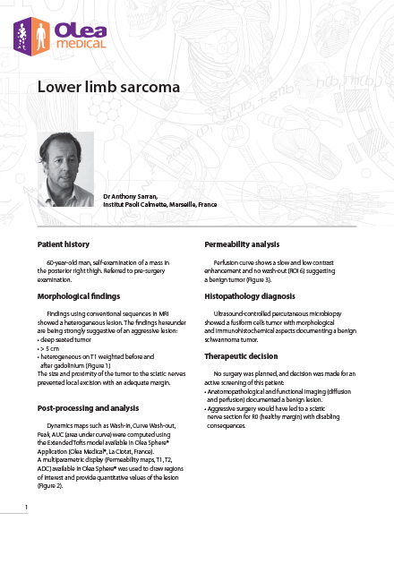
    60-year-old man, self-examination of a mass in the posterior right thigh. Referred to pre-surgery examination.

Overview of the main functions

Automatic calculation of ADC and synthetic b maps
Automatic calculation of multi-parameter T1 perfusion maps
Automatic analysis of the kinetic enhancement curves
Quantitative analysis of the relaxation time
Quantification of the lipid and iron fractions for the analysis of metabolic disorders
Customizable multi-parameter display and 3D rendering
Measurement calculation
Segmentation of the volume of interest
Standardized reports based on the Bi-RADS® (breast) and PI-RADS® (prostate) classifications
Texture  Optional

Give it a go!

Request a demo or contact our sales team

Renseignez votre email dans le formulaire ci-dessous afin de pouvoir télécharger les case reports sur le site olea-medical